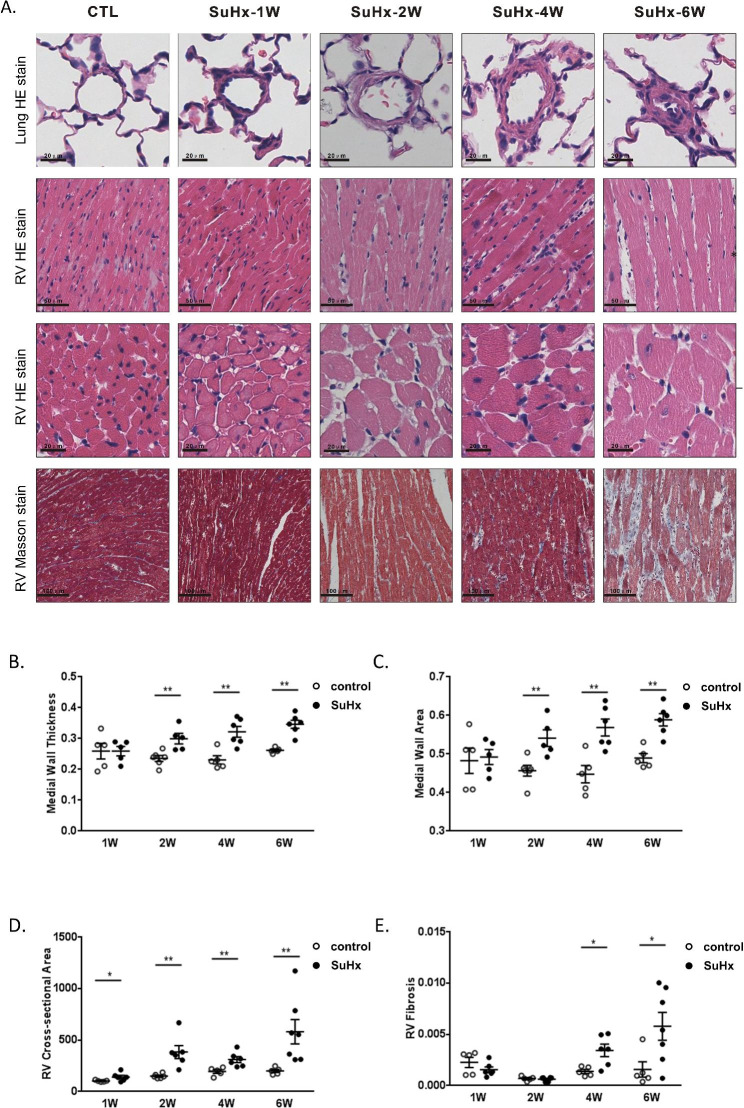
Fig. 2.
Changes of pulmonary vascular medial wall thickness and RV lesions in SuHx rats. (A) Representative photomicrographs of hematoxylin and eosin (H&E) stain of pulmonary vessel cross sections, H&E stained and masson’s trichrome stain of RV sections. (B, C) Scatter dot plot showing the comparison of vessel wall thickness and vessel wall area of vessels (outside diameter > 50 μm) between the CTL and SuHx groups. (D, E) Scatter dot plot showing the comparison of cross-sectional area of right ventricle cardiomyocyte and fibrosis between two groups. Data were presented as mean ± S.E.M., control group n = 5–7, SuHx group n = 6–7, *P < 0.05, **P < 0.01, compared with the control group. W: week

