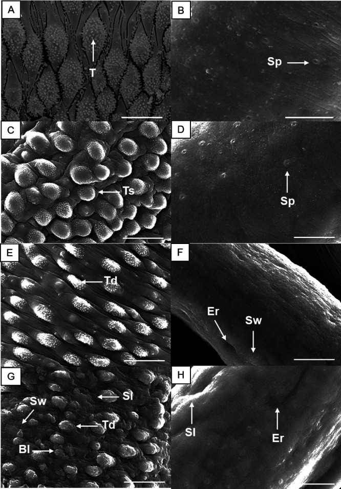
FIG 2.

Scanning electron microscopy of adult S. mansoni following incubation with nifuroxazide (NFZ). Control male parasite showing intact tubercles (T) and spines on the surface (A), and control female worm (B) showing the sensory papillae (Sp). Schistosomes were exposed to NFZ at 12.5 μM (C and D), 25 μM (E and F), and 50 μM (G and H). Male (A, C, E, and G) and female (B, D, F, and H) schistosomes. The dorsal tegumental surface shows tubercle shortening (Ts), swelling (Sw), erosion (Er), sloughing (Sl), blisters (Bl), and tubercle disintegration (Td). Images were captured using a JEOL SM 6460LV scanning electron microscope after 72 h of incubation. Scale bars: 10 μm.
