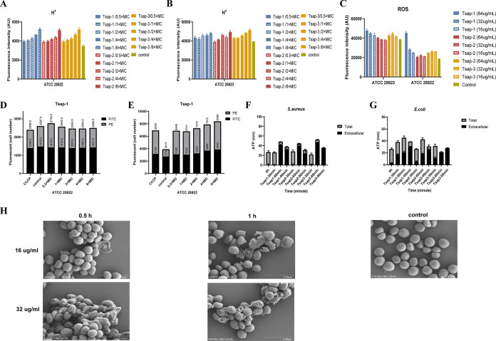
FIG 6.
Tsap peptides disrupt the membranes of E. coli and S. aureus. The H+ content of ATCC 25922 (A) and ATCC 25923 (B) after treatment with Tsap peptides at various concentrations is shown. Untreated bacteria were used as a control. (C) ROS levels in ATCC 25922 and ATCC 25923 after treatment with Tsap AMPs at various concentrations. The effect of Tsap on membrane permeability in Escherichia coli (D) and Staphylococcus aureus (E) is shown. FITC staining and flow cytometry were used to determine the number of bacteria with altered membrane potential (PE) and the number of unaltered bacteria after exposure to Tsap-1 at 0.5× MIC, 1× MIC, 2× MIC, 4× MIC, and 8× MIC. The CCCP-treated group was used as a positive control. Determination of ATP leakage in S. aureus (F) and E. coli (G) treated with AMPs for different amounts of time is shown. The bacterial cells were treated with Tsap AMPs at 1× MIC for 30 min, 60 min, and 90 min, and intracellular and extracellular ATP contents were then determined. (H) Shows scanning microscopy images of Tsap-1-treated S. aureus. Scanning microscopy images of S. aureus ATCC 25923 are also shown after treatment with Tsap-1 at 16 μg/mL or 32 μg/mL for 0.5 h or 1 h, respectively. Untreated ATCC 25923 were used as a negative control.

