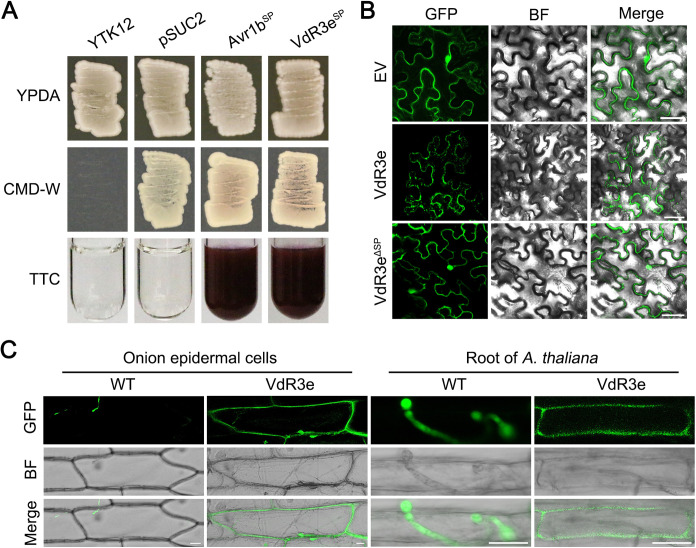
FIG 3.
Subcellular localization of the V. dahliae VdR3e in plant tissues. (A) Validation of the signal peptide activity of VdR3e by yeast signal trap assay. The yeast strain YTK12 can grow on yeast extract peptone dextrose adenine (YPDA) medium and YTK12 containing the pSUC2 vector can grow on CMD-W medium. The fusion of VdR3e signal peptide with mature yeast invertase enables the invertase to be secreted. As a result, the color of TTC solution changes from colorless to red. Avr1b was used as a positive control. (B) Subcellular localization of VdR3e-GFP in N. benthamiana leaves. VdR3e and VdR3eΔSP were transiently expressed in N. benthamiana leaves by agroinfiltration. The pBin-GFP was used as a negative control. The fluorescence was scanned by Lecia TCS SP8 confocal microscopy system with an excitation wavelength at 488 nm and emission wavelength at 510 nm. Scale bars, 50 μm. (C) Subcellular localization of VdR3e-GFP in onion epidermal cells and roots of A. thaliana. The onion epidermis and roots of A. thaliana were immersed in the conidial suspension of HoMCLT overexpressing VdR3e-GFP and GFP for 20 min. The onion epidermal or A. thaliana tissues were incubated on water agar medium for 5 and 2 days, respectively. Finally, fluorescence was observed with confocal microscopy. Scale bars, 25 μm.

