FIG 3.
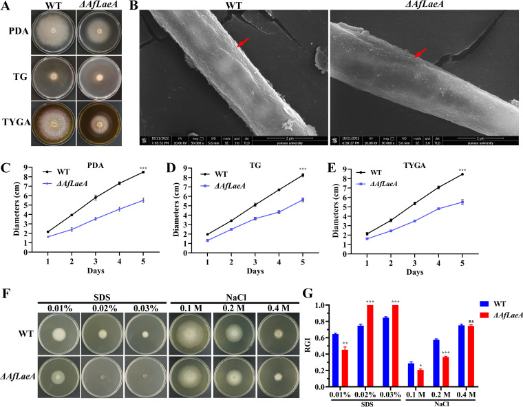
Comparison of growth, morphologies, and stress responses between WT and ΔAfLaeA strains. (A) Colony morphologies of the WT and ΔAfLaeA strains incubated on PDA, TG, and TYGA plates at 28°C for 5 days. (B) Analysis of the cell surface morphology of WT and ΔAfLaeA strains by SEM. Red arrows indicate wrinkled areas on the hyphal surface. (C to E) Colony diameters of the WT and ΔAfLaeA strains cultured on PDA, TG, and TYGA plates at 28°C for 5 days (***, P < 0.001). (F) Growth of the WT and ΔAfLaeA strains on medium supplemented with SDS at 0.01% to 0.03% and NaCl (0.1 M, 0.2 M, and 0.4 M). (G) RGI values of the WT and ΔAfLaeA strains on medium supplemented with SDS (0.01% to 0.03%) and NaCl (0.1, 0.2, and 0.4 M) (*, P < 0.05; **, P < 0.01; ***, P < 0.001).

