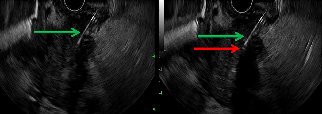
Figure 2.
EUS-guided placement of a diffusing fiber for delivery of PDT. A, The 19-gauge FNA needle (green arrow) is visualized within the pancreatic head mass under endosonography. B, The diffusing tip of the optical fiber is seen after introduction through the needle under endosonography as a small hyperechoic point (red arrow).

