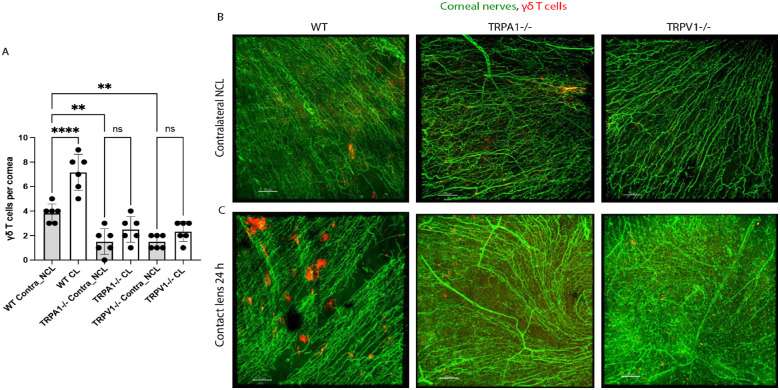
Figure 3.
Quantification (A) and representative immunofluorescence imaging (B, C) of γδ T cells (red) in the corneas of wild-type or gene-knockout mice in TRPA1 (−/−) or TRPV1 (−/−) with or without contact lens wear for 24 hours. Corneal nerves were labeled with β-tubulin III (green). Scale bar = 70 µm. The γδ T cell responses to lens wear (white bars) were absent in both TRPA1 (−/−) and TRPV1 (−/−) mice. Contralateral eyes of TRPA1 (−/−) and TRPV1 (−/−) mice (grey bars) each also showed a significant reduction in baseline numbers of γδ T cells. ** P < 0.01, **** P < 0.0001, ns = not significant [One-way ANOVA with Tukey's multiple comparisons test].

