Figure 5.
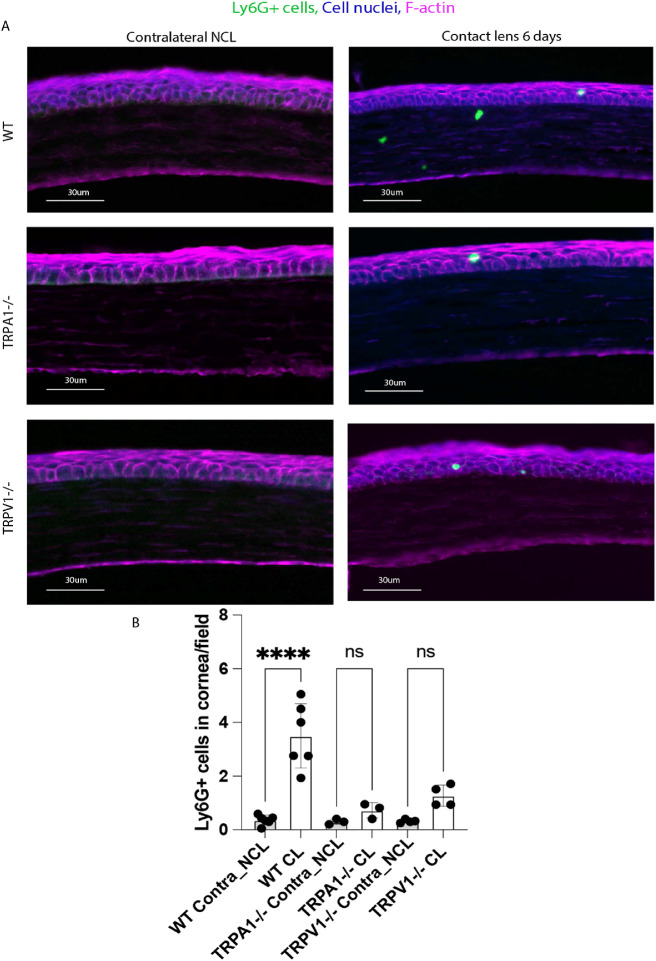
(A) Immunofluorescence imaging of corneal cryosections showing Ly6G+ cell (neutrophil) infiltration of a mouse cornea after 6 days of contact lens wear versus the no lens wear contralateral eye. Lens-induced Ly6G+ cell infiltration was reduced in the corneas of lens wearing TRPA1 (−/−) and TRPV1 (−/−) mice. Ly6G+ cells (green), cell nuclei (blue), and cell F-actin (red). Scale bar = 30 µm. (B) Quantification of Ly6G+ cells (per field of view) in WT versus TRPA1 (−/−) and TRPV1 (−/−) corneas after 6 days of contact lens wear versus contralateral eyes. The lens-induced Ly6G+ response was lost in both TRPA1 (−/−) and TRPV1 (−/−) mice. **** P < 0.0001, ns = not significant [One-way ANOVA with Tukey's multiple comparisons test].

