. 2023 Aug 17;12:RP87283. doi: 10.7554/eLife.87283

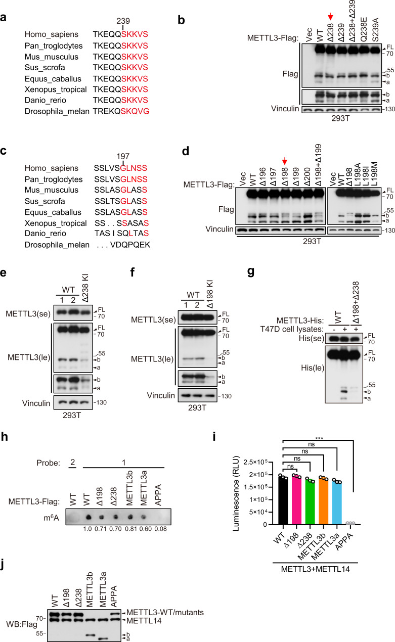
Figure 2. METTL3a (residues 239–580) and METTL3b (residues 197–580) are post-translational products through highly conserved residues.

Sequence alignments of the conserved residues on (234–244) (a) and (192–201) (c) of METTL3. (b, d) Immunoblot of 293T cells transfected with empty vector (Vec) or METTL3-WT (wild-type) or the indicated mutants. Immunoblot analysis of 293T cells with CRISPR knock-in (KI) mediated deletion of Q238 (e) or L198 (f). (g) Purified recombinant METTL3-WT-His or METTL3-(Δ198+Δ238)-His protein were incubated with or without T47D cell lysates at 37°C for 1 hr followed by immunoblot with anti-His antibody. (h–j) The in vitro protein methylation activity was tested using purified METTL3-WT-Flag and its mutant proteins in combination with co-purified Flag-METTL14 and RNA-probe. The methylation activity was measured by using dot blot (h) or the Promega bioluminescence assay (i), and the immunoblot of those purified proteins are shown in (j). Error bars represent mean ± standard deviation (SD), unpaired t-test. ***p < 0.001, ns denotes no significance. FL indicates the full-length of METTL3. The short forms are labeled as a and b. se and le indicated short exposure and long exposure, respectively.

Figure 2—source data 1. Unedited western blot images for Figure 2.
Figure 2—source data 2. Table related to Figure 2i.

Figure 2.

Figure 2—figure supplement 1. Distribution of METTL3-WT and its mutants in breast cancer cells.
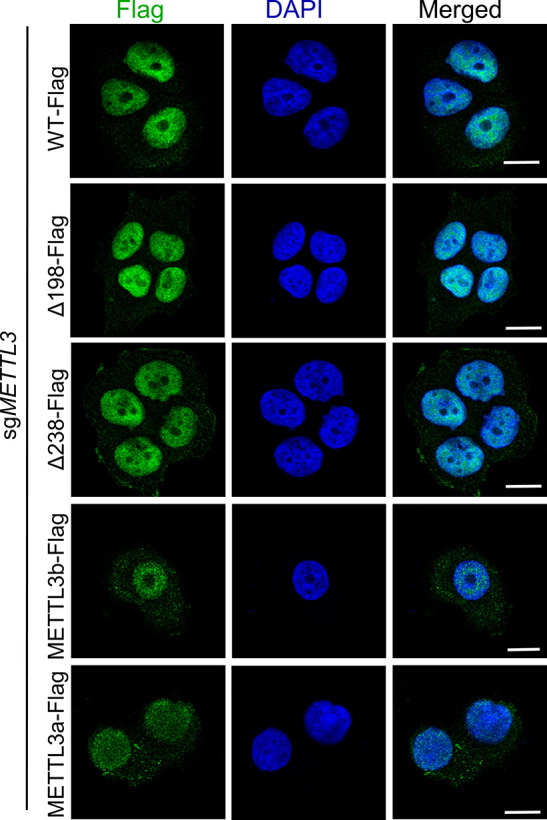
Figure 2—figure supplement 1.

Immunofluorescence analysis of T47D cells stably expressing METTL3-WT or mutants under METTL3 knockout. Scale bars, 10 μm.
Figure 2—figure supplement 1—source data 1. Images related to Figure 2—figure supplement 1.
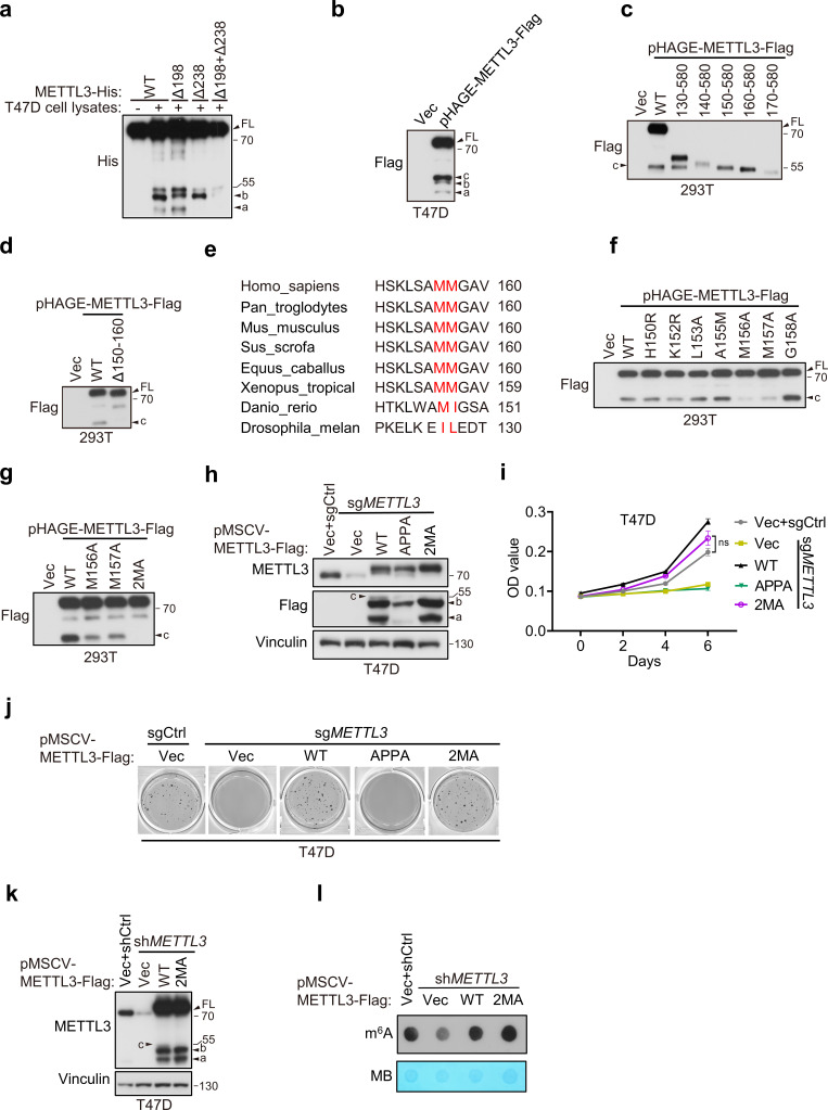
Figure 2—figure supplement 2. The short form METTL3c is dispensable for METTL3-mediated functions.

Figure 2—figure supplement 2.

(a) Purified recombinant METTL3-WT-His, METTL3-Δ198, METTL3-Δ238, or METTL3-(Δ198+Δ238)-His protein were incubated with or without T47D cell lysates at 37°C for 1 hr followed by immunoblot with anti-His antibody. (b) Immunoblot of T47D cells infected with pHAGE-METTL3-Flag. (c, d, f, g) Immunoblot of 293T cells transfected with pHAGE-METTL3-WT or its indicated mutants. (e) Sequence alignment of conserved amino acids on (150–160) of METTL3. Immunoblot analysis (h), cell proliferation (i), and soft agar growth (j) of T47D cells infected with retrovirus encoding Vector (Vec), sgMETTL3 resistant METTL3-WT or its indicated mutants followed by another infection with sgMETTL3 or sgControl (sgCtrl). Immunoblot analysis (k) and N6-methyladenosine (m6A) dot blot (l) in 293T cells infected with shMETTL3 or shControl (shCtrl) followed by transfection with indicated plasmids. Methylene blue (MB) was used as a loading control. Error bars represent mean ± standard error of the mean (SEM), unpaired t-test. ns denotes not significant. FL is the full-length of METTL3. The short forms are labeled a, b, and c.
Figure 2—figure supplement 2—source data 1. Unedited western blot images for Figure 2—figure supplement 2a–d, f–h, and j–l.
Figure 2—figure supplement 2—source data 2. Table related to Figure 2—figure supplement 2i.