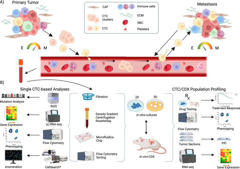
Fig. 1. Circulating tumor cells (CTCs) leave the primary tumor as single cells or in clusters, intravasate into the bloodstream and travel through the circulation to the distant site of the body to establish metastasis.
A At the primary site, epithelial-to-mesenchymal transition (EMT) leading to more mesenchymal (M) phenotype is characteristic for CTCs, whereas in metastatic site, more epithelial (E) phenotype and mesenchymal-to-epithelial transition occurs. Moreover, cancer cells can leave the metastatic site and colonize back its primary tumor site (tumor self-seeding). B For establishment of in vivo/in vitro preclinical models, viable CTCs are isolated/enriched from peripheral whole blood by several methods (filtration, density gradient centrifugation coupled with negative depletion, microfluidic devices, or flow cytometry). Obtained CTCs can be characterized using various single cell-based technologies or used for establishment of in vitro culture (suspension/adherent/tumoroids) and in vivo CTC-derived xenografts (CDX). In vitro cultures may be used for establishment of CDX and vice versa, developed CDX may serve as a source of material for establishment of in vitro cultures. Both in vivo/in vitro models serve as a source of valuable material for subsequent analyses of CTCs at the single cell level (mutation analyses, sc-RNA-Seq, mass cytometry, microscopy), as well as analysis of bulk population (drug screening and therapy decision making, phenotype analysis using flow cytometry). CAF cancer-associated fibroblasts, RBC red blood cells, ECM extracellular matrix. Created with BioRender.com.

