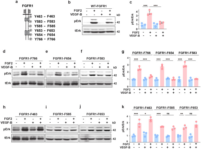
Fig. 4.
FGFR1 tyrosine residues important for the inhibitory effect of VEGF-B on FGF2-induced Erk activation. a Scheme showing the tyrosine (Y) residues of FGFR1 replaced by phenylalanine (F). b–g Western blots showing that VEGF-B (100 ng/ml, 30 min treatment) inhibits FGF2 (50 ng/ml)-induced Erk phosphorylation in cells expressing wild-type FGFR1 (WT-FGFR1, b, c) or the FGFR1-F766 (d, g), FGFR1-F654 (e, g), FGFR1-F583 (f, g) mutants. n = 3 each group. For (c), adjusted p values are 9.7E−5 for FGF2 vs. BSA and 3.4E−4 for FGF2 vs FGF2 + VEGF-B. For FGFR1-F766, FGFR1-F654, and FGFR1-F583 in (g), adjusted p values are 4.0E−8 for BSA vs FGF2; 7.4E−6 for FGF2 vs FGF2 + VEGF-B; 1.2E−5 for FGF2 vs. BSA; 1.4E−5 for FGF2 vs FGF2 + VEGF-B; 2.9E−5 for FGF2 vs. BSA and 3.6E−5 for FGF2 vs FGF2 + VEGF-B. h–k Western blots showing that in cells expressing the FGFR1-F463 (h, k), FGFR1-F585 (i, k), or FGFR1-F653 (j, k) mutants, VEGF-B failed to inhibit FGF2-induced Erk phosphorylation. Adjusted p values are 6.0E−8 for FGFR1-F463; 1.2E−5 for FGFR1-F585 and 1.3E−3 for FGFR1-F653. For (c), (g), and (k), one-way ANOVA followed by Sidak post hoc analysis was used (number of comparisons is 2 for c, g, and k). All data are mean ± s.e.m. *p < 0.05, **p < 0.01, ***p < 0.001, ns: p > 0.05. The experiments were repeated three times

