FIGURE 2.

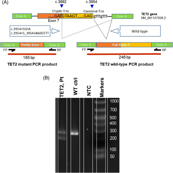
(A) Graphic representation showing TET2 splicing products with or without TET2 intronic variants (c.3954+5_3954+8delGTTT, c.3954+5G>A). Breakpoints at the cryptic and original 5′ splice site junctions are shown with blue triangles. The primer binding sites and subsequent PCR products in WT and mutant TET2 spliced mRNA are also shown. (B) RT‐PCR analysis on RNA from a negative control patient and a patient with TET2 c.3954+5_3954+8del variant. Gel electrophoresis image showing PCR products amplified using specific primers binding to Ex6 (forward) and Ex8 (reverse). Fragment size of 246 bp indicates TET2 wild‐type sequences (ex6+ex7+ex8) and fragment of 185 bp size (ex6+ partial ex7+ex8) indicates the TET2 splicing variant. Only wild‐type product was identified in the negative (WT) control. No visible product is shown in no template control (NTC).
