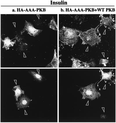
FIG. 6.
Coexpression of wild-type PKB with AAA-PKB rescues the inhibition of insulin-stimulated GLUT4 translocation. L6-GLUT4myc myoblasts were cotransfected with GFP (0.4 μg), HA-AAA-PKB (0.4 μg), and pcDNA3 (1.6 μg) (a) or with GFP (0.4 μg), HA-AAA-PKB (0.4 μg), and HA-PKB (1.6 μg) (b) and incubated for 48 h in culture. Cells were serum deprived for 5 h, left untreated or treated with 100 nM insulin for 20 min, and then processed for cell surface GLUT4myc detection as indicated in Materials and Methods. Untreated cells are not shown. Each pair of panels (upper and lower) shows the same field of cells. In the lower panels, GFP fluorescence in transfected cells is shown (arrowheads). The upper panels show the cell surface GLUT4myc density for the transfection of AAA-PKB plus pcDNA3 vector alone (a) or the transfection of AAA-PKB with excess wild-type (WT) PKB (b). Arrowheads indicate the positions of transfected cells. Results shown are representative of at least three experiments.

