Figure 2.

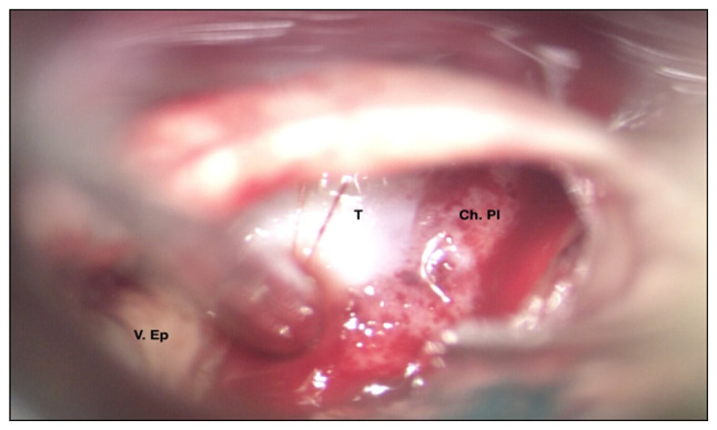
Intraoperative photo of the tumour, appearing as a whitish lesion with vascularised areas and a hard-friable consistency. T, tumour; V. Ep., epidural vein; Ch. Pl., choroid plexus.

Intraoperative photo of the tumour, appearing as a whitish lesion with vascularised areas and a hard-friable consistency. T, tumour; V. Ep., epidural vein; Ch. Pl., choroid plexus.