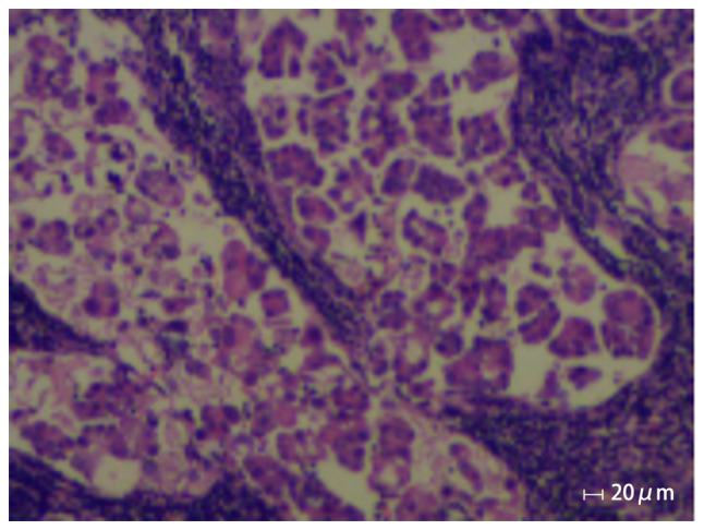
Figure 1.

Histopathology showing cancer cells in the lymphoid tissue from the right supraclavicular fossa since the first visit in April 2016. Epithelioid cells are present in scattered nests and micropapillary structures, and focal calcifications, cytological atypia and mitotic figures occur in the lymphoid tissue from the right supraclavicular fossa.
