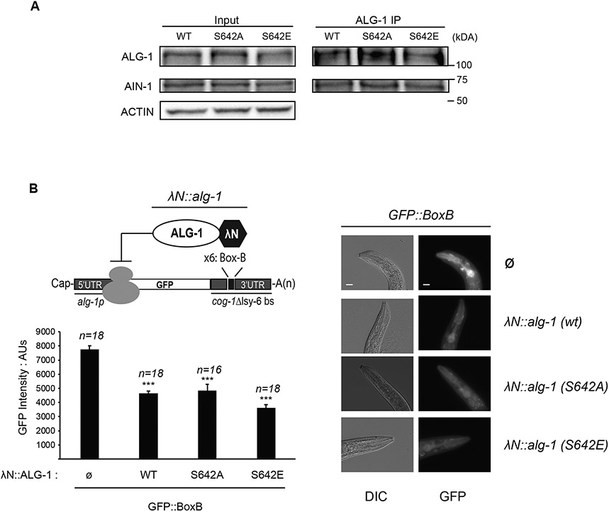
Figure 3. Phosphorylation of ALG-1 serine 642 does not affect its interaction with GW182 homolog AIN-1 nor its ability to silence a mRNA.
(A) ALG-1 co-immunoprecipitation with GW182 homolog AIN-1. ALG-1 was immunoprecipitated from adult extract of wild type, alg-1(S642A) or alg-1(S642E) and polyclonal antibodies for AIN-1 and ALG-1 were used for western blotting. The inputs are 10% of the total protein extracts used for immunoprecipitations. Actin served as loading control. The blots are representative of three biological replicates. (B) ALG-1 S642 phosphorylation does not impair mRNA silencing when tethered to a mRNA 3′ UTR. Top left: Schematic representation of AGO tethering system. A GFP reporter under the control of an alg-1 promoter fused with the sequence of cog-1 3′UTR where the lsy-6 miRNA binding sites (delta lsy-6 bs) are replaced by six copies of the Box-B element (x6: Box-B). The high affinity between the Box-B RNA secondary structure and the λN peptide fused to ALG-1 leads to its recruitment. A strain with a single integrated copy of alg-1p::GFP::Box-B reporter carrying endogenous alg-1 alleles tagged with a λN sequence at the 5′ end of the coding sequence was used to edit λN::alg-1 into non-phosphorylatable λN::alg-1 (S642A) and phospho-mimicking λN::alg-1 (S642E). The expression level of the GFP reporter was measured in the pharynx. Bottom left: The GFP level expressed in the pharynx of young adult worms was quantified using arbitrary units (AU). The error bars represent the 95% confidence interval, and the P-values indicated were measured by a two-tailed Student’s t-test; *** p<0.001. The number of animals scored (n=) is indicated and the graph is representative of two independent measurements. Right: Representative images of animals expressing only the GFP reporter (Ø) or GFP reporter and different versions of λN-tagged alg-1 (λN::alg-1) gene are shown. The scale bar indicates 20 μm. Images were obtained at the same time of exposure, on the same slide, and with the same area of measure for each animal strain.

