Fig. 3.
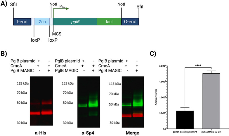
Designing and testing of MAGIC v.2; A schematic diagram of I and O end of MAGIC v.2; B Western blot of 5 µg His-tagged CmeA protein purified by nickel affinity chromatography. Biological samples were separated on a Bolt 4–12% bis–tris gel (Invitrogen) with MOPS buffer and transferred to nitrocellulose membrane with an iBlot 2 dry blotting system. The membrane was probed with anti-His (Invitrogen) and anti-SP4 (Statens, Serum Institute) and detected with fluorescently labelled secondary antisera (red-His, green- anti-SP4) on a LI-COR Odyssey scanner.; C densitometry analysis of glycoconjugate production in E. coli MAGIC v.2 SP4 compared to E. coli bioconjugation SP4.; Densitometry analysis of glycoconjugate was done from three biological replicates. Statistical analysis is from three biological replicates using Student’s t-test ns, p > 0.05; *,p < 0.05, **,p < 0.01, ***, p < 0.001

