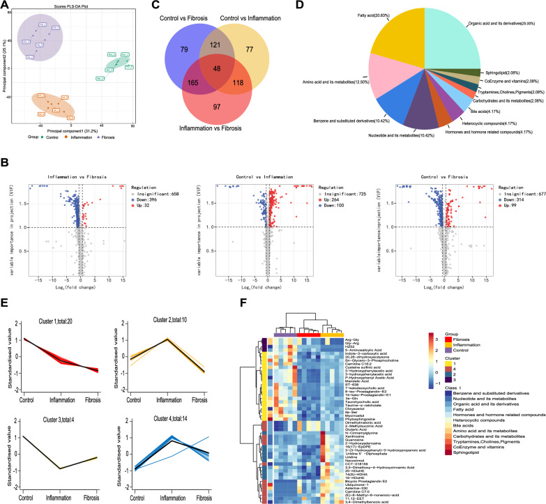
Fig. 3.
Altered metabolic profiles in the feces of the fibrosis mouse model. A PLS-DA multivariate statistical model of the Control, Inflammation, and Fibrosis groups. B Volcano plot of metabolites, with blue representing down-regulation, gray representing non-significance, and red representing up-regulation. C Venn diagram shows the shared or unique differential metabolites among the three groups. D Classification of the 48 SAMs among the three groups. Each color displays a class of metabolites, with the specific percentage highlighted in the pie chart. E K-means clustering diagram of the 48 SAMs among the three groups, with the specific number of metabolites presented in each cluster, and all the clusters follow the timeline of Control (Ctrl), Inflammation (TNBS-4W), and Fibrosis (TNBS-6W). F Cluster heatmap of the relative abundance of the 48 SAMs. n = 5 per group. PLS-DA partial least squares discriminant analysis, SAMs significantly altered metabolites

