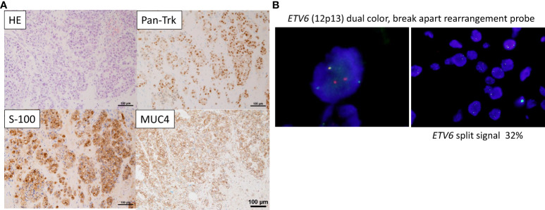
Figure 1.
Pathological findings (A) On Hematoxylin & Eosin (HE) staining, tumor cells with well-defined nucleoli and a pale eosinophilic, partially vacuolated cytoplasm resemble a microcystic form, presenting a histology similar to the microcystic form of acinic cell carcinoma. Immunostaining showing that Pan-Trk and S-100, and MUC4 are widely expressed. (B) Fluorescence in situ hybridization (FISH) using the ETV6 break apart probe examined for the presence of yellow (red/green fusion) or green/red fluorescent signals in the tumor cells. Yellow signals are negative, and separate red and green signals are positive. ETV6 split signal of 32% is seen (cutoff, 10%).

