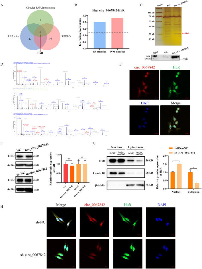
Fig. 4.
Hsa_circ_0067842 promotes the translocation of HuR protein into the cytoplasm. A Venn diagram showing the RBPs interacted with hsa_circ_0067842. B Interaction probabilities between HuR and hsa_circ_0067842 predicted by RPISeq (> 0.5 were considered “positive”). C Western blot of the proteins from hsa_circ_0067842 pull-down assay, followed by silver staining. D Three HuR protein-specific peptides were identified by mass spectrometry. E FISH for hsa_circ_0067842 (red) and HuR (green) in BC cells (MDA-MB-468 cell line). DAPI were used to stained for nuclei (blue). Scale bars are 20 μm. F The relative expression of HuR was detected by western blot after hsa_circ_0067842 overexpression (MCF-7) or knockdown (MDA-MB-468). G After knocking down hsa_circ_0067842, the level of nuclear HuR protein significantly increased, while the cytoplasmic HuR protein obviously decreased. H Fluorescence localization analysis of hsa_circ_0067842 and HuR showed that downregulating hsa_circ_0067842 could inhibit the translocation of HuR into the cytoplasm. The data are assessed as the mean ± SD, nsp > 0.05, *p < 0.05, and ***p < 0.001

