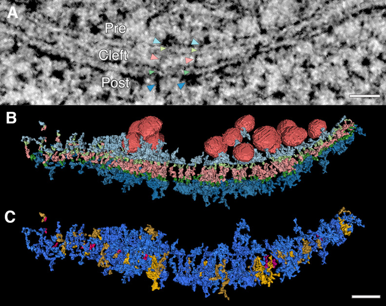
Figure 1.
Electron tomography reveals extensive transsynaptic connection of intracellular structures throughout the synapse. Transcleft, transmembrane, and intracellular electron-dense structures were hand segmented and rendered in 3D from electron tomograms of high-pressure frozen and freeze-substituted mature dissociated rat hippocampal neuronal cultures. A, |Transsynaptic assemblies, or assemblies of connected electron-dense material (black pixels) that span the synapse, are found in 15-nm virtual projections, as seen here. Color coded arrowheads indicate structures of two transsynaptic assemblies. Light blue arrowheads point to presynaptic (pre) membrane-bound structures. Light green arrowheads point to presynaptic transmembrane structures. Pink arrowheads point to transcleft (cleft) structures. Dark green arrowheads point to postsynaptic transmembrane structures. Dark blue arrowheads point to postsynaptic (post) membrane-bound structures. B, A single synapse is rendered with synaptic vesicles (red) associated with transsynaptic assembly and all assembly structures (color coded the same as arrowheads in A to indicate the abundance of structures on both sides of the synapse connecting to transcleft structures. C, Transsynaptic assemblies further classified with a different color code. An assembly with no intracellular structure is considered transcleft only (pink); an assembly that contains a transcleft structure connect to an intracellular structure on one side of the synapse is classified as a partial assembly (yellow); and an assembly with a transcleft structure connected to intracellular structures on both sides is classified as full transsynaptic assembly (blue). Scale bars: 40 nm.

