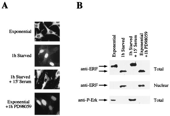
FIG. 4.
Phosphorylation-dependent subcellular localization of ERF. (A) Ref-1 cells growing under the indicated conditions were fixed and stained with the S17S anti-ERF specific antibody and visualized by fluorescence microscopy (magnification, ×70). (B) Total and nuclear protein extracts from the same cells were analyzed by immunoblotting with the same anti-ERF specific antibody. Activated Erks were detected with an antibody directed against their phosphorylated form.

