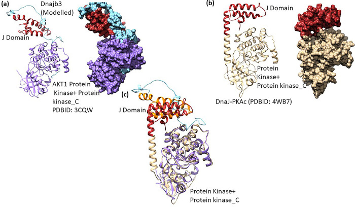
Fig 4. Docking of modelled DNAJB3 onto AKT1 and structural comparison.
(a) Molecular docking of modelled DNAJB3 (J domain: red, c-terminal: sea green) onto AKT1 (PDBID:3CQW, purple) represented as cartoon (left panel) and surface (right panel). (b) Structure of Dnaj-PKACα (PDBID: 4WB7) drawn in carton (left panel) and surface (right panel) representations. The PKACα is coloured golden and J domain as red. (c) Structure alignment of docked (DNAJB3: AKT1) structure to Dnaj-PKACα (PDBID: 4WB7).

