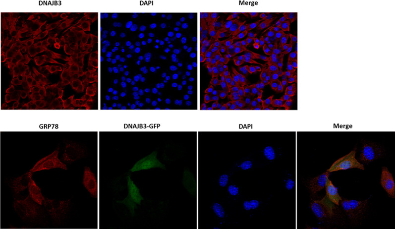
Fig 5. DNAJB3 sub-cellular localization with ER marker using confocal microscopy.
Staining with anti-DNAJB3 in C2C12 cells reveals its non-nuclear localization (upper panel). C2C12 cells were transfected with DNAJB3 tagged with GFP (DNAJB3-GFP) and stained with anti-GRP78 (an ER marker) and found that they colocalize (lower panel). DAPI was used as control for nuclear staining.

