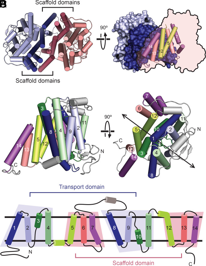
Fig. 2.
Fold and oligomeric structure of PurTCp. (A) The PurTCp dimer viewed from the extracellular side, with the scaffold and transport domains colored in lighter or darker shades, respectively. (B) A view of the dimer from within the plane of the membrane. One protomer is shown with a surface view, and the other is shown as a transparent outline with helices from the scaffold domain shown as cylinders. (C) Two perpendicular views of a protomer from the PurTCp structure colored in pairs of symmetry-equivalent transmembrane helices. The twofold pseudosymmetry axis is marked on the right. (D) A topology diagram colored according to the same scheme as in panel C.

