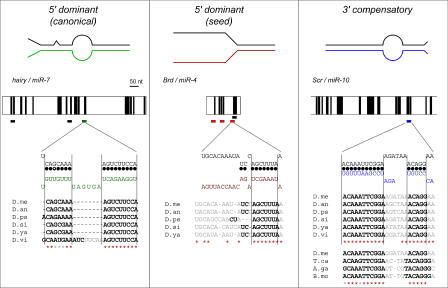
Figure 4. Three Classes of miRNA Target Sites.
Model of canonical (left), seed (middle) and 3′ compensatory (right) target sites. The upper diagram illustrates the mode of pairing between target site (upper line) and miRNA (lower line, color). Next down in each column are diagrams of the pattern of 3′ UTR conservation. The vertical black bars show stretches of at least six nucleotides that are conserved in several drosophilid genomes. Target sites for miR-7, miR-4, and miR-10 are shown as colored horizontal bars beneath the UTR. Sites for other miRNAs are shown as black bars. Furthest down in each column the predicted structure of the duplex between the miRNA and its target site is shown; canonical base-pairs are marked with filled circles, G:U base-pairs with open circles. The sequence alignments show nucleotide conservation of these target sites in the different drosophilid species Nucleotides predicted to pair to the miRNA are shown in bold; nucleotides predicted to be unpaired are grey. Red asterisks indicate 100% sequence conservation; grey asterisks indicate conservation of base-pairing to the miRNA including G:U pairs. The additional sequence alignment for the miR-10 target site in Scr in Tribolium castanaeum, Anopheles gambiae, and Bombyx mori strengthens this prediction. Note that the reduced quality of 3′ compensation in these species is compensated by the presence of a better quality 7mer seed. A. ga, Anopheles gambiae; B. mo, B. mori; D. an, D. ananassae; D. me, D. melanogaster; D. ps, D. pseudoobscura; D. si, D. simulans; D. vi, D. virilis; D. ya, D. yakuba; T. ca, T. castanaeum.

