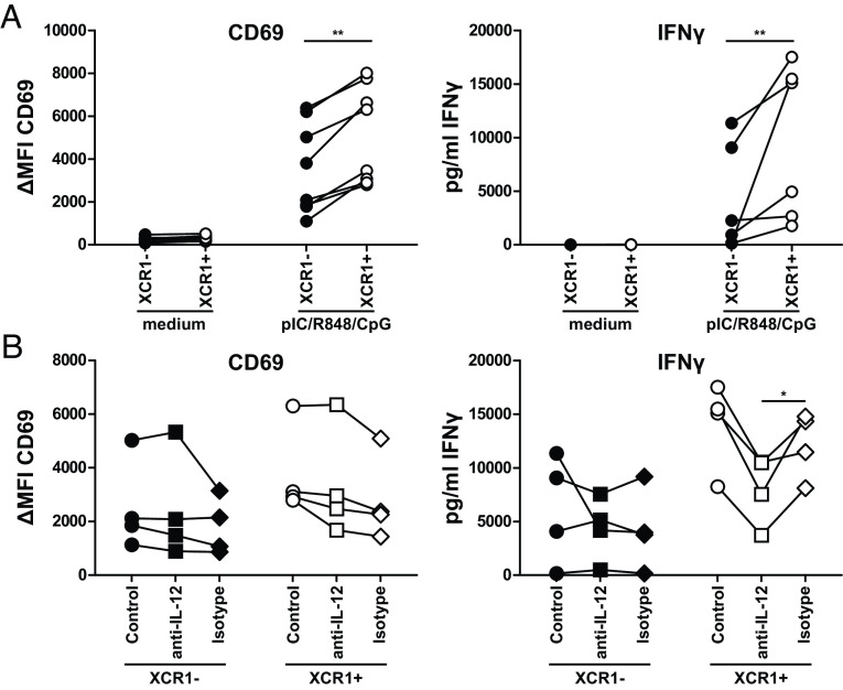
Fig. 4.
XCR1+ cDC1 excel in the activation of NK cells due to IL-12 secretion. Sorted XCR1− or XCR1+ cDC1 were cocultured with NK cells in a 1:5 ratio in presence or not of 2.5 µg/mL R848, 2.5 µg/mL pIC and 2.5 µg/mL CpG (Type A) as described in SI Appendix, Fig. S10A. After 18 h, supernatants were harvested and analyzed for the secretion of IFNγ by CBA Flex-Set (BD Biosciences) as well as NK cells for the expression of CD69 by flow cytometry. Samples were acquired using a BD LSRFortessa and analyzed using FlowJo (CD69 expression) or FCAP Array 3.1 (IFNγ secretion). (A) Scatter plots show ΔMFI for CD69 (N = 8, each donor connected by line) and secretion of IFNγ (N = 6, each donor connected by line). (B) Assay was performed as described in (A) with the addition of 10 µg/mL of a neutralizing anti-IL-12/IL-23p40 antibody (clone: C11.5) or an appropriate isotype control during the 18 h coculture. Scatter plots show ΔMFI for CD69 (N = 4, each donor connected by line) and secretion of IFNγ (N = 4, each donor connected by line). *P < 0.05; **P < 0.01 (paired Student’s t test).

