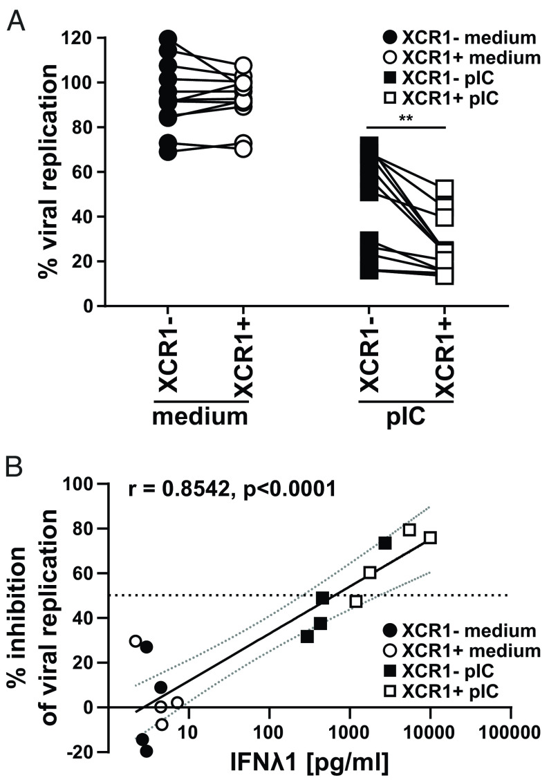
Fig. 5.
XCR1+ cDC1 are superior in the induction of an antiviral state in epithelial cells. Sorted XCR1− and XCR1+ cDC1 were stimulated or not with 5 µg/mL pIC as described in SI Appendix, Fig. S10B Supernatants (SN) were harvested and used for the pretreatment of U2OS cells. After overnight culture, U2OS cells were challenged with luciferase-expressing Influenza virus type A (strain PR8-Luc). (A) Scatter plots show viral replication (% compared to nontreated infected cells) in U2OS cells after treatment with SN of XCR1− and XCR1+ cDCs ± stimulation with 5 µg/mL pIC. XCR1− (filled black circles: medium-treated; filled squares: pIC-treated) and XCR1+ cDC1 (open circles: medium-treated; open squares: pIC-treated) from the same donor are connected by lines (N = 12; **P < 0.01; paired Student’s t test). (B) SN from four donors were used for pretreatment of U2OS cells as in (A) and analyzed for the secretion of IFNλ1 by CBA assay (LEGENDplex Human Anti-Virus Response Panel). Scatter plots show inhibition of viral replication in U2OS cells correlated with the amount of IFNλ1 in the used SN for pretreatment (N = 4; XCR1− cDC1: black symbols, XCR1+ cDC1: white symbols; control-treated: circles, pIC-treated: squares, horizontal line depicts 50% inhibition of viral replication). Semilog line was fitted and Pearson Correlation Coefficient r and P value (two-tailed, exact) determined using GraphPad Prism9.

