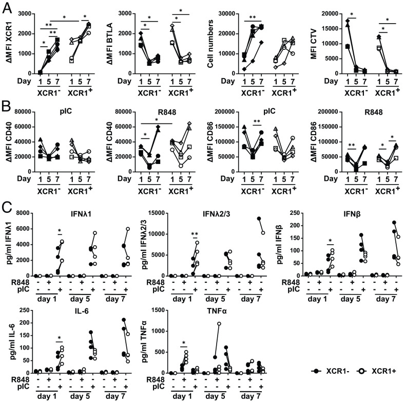
Fig. 6.
Differentiation of XCR1− cDC1 into XCR1+ cDC1 enables secretion of inflammatory cytokines. Sorted XCR1− and XCR1+ cDC1 were cultured in a DC differentiation assay as described in SI Appendix, Fig. S10C. On days 0, 4, and 6, cells were stimulated for 12 h or not with either 5 µg/mL pIC or R848. SN were harvested and cells were analyzed by flow cytometry for XCR1, BTLA, CD40, and CD86 expression. SN were analyzed for cytokine and chemokine secretion by CBA assay (LEGENDplex Human Anti-Virus Response Panel). (A) Scatter plots show XCR1 and BTLA expression (ΔMFI values), cell numbers as well as MFI value of CTV signal and (B) CD40 and CD86 expression (ΔMFI values) (filled black symbols: XCR1− cDC1; open symbols: XCR1+ cDC1; each individual donor has a distinct symbol and is connected by lines; N = 4; *P < 0.05, **P < 0.01; Two-way ANOVA). (C) Harvested SN of the samples in (B and C) were analyzed for the secretion of IFNλ1, IFNλ2/3, IL-6, TNFα, and IFNβ secretion by CBA assay (LEGENDplex Human Anti-Virus Response Panel). Concentration of the cytokines is shown as scatter plots (filled black circles: XCR1− cDC1; open circles: XCR1+ cDC1; individual donors connected by lines; N = 4; *P < 0.05, **P < 0.01; paired Student’s t test).

