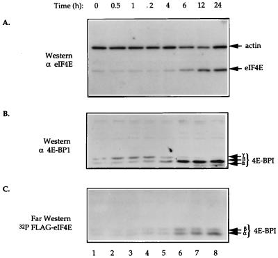
FIG. 5.
eIF4E induction results in dephosphorylation of 4E-BP1. Cell extracts were prepared from C1 cells in the absence of tetracycline for the indicated times. (A) Total cell extract (25 μg) was resolved on an SDS–12.5% polyacrylamide gel, electroblotted onto a 0.45-μm-pore-size nitrocellulose membrane, and probed with rabbit polyclonal anti-eIF4E and mouse monoclonal anti-actin antibodies. (B) Total cell extract (50 μg) was resolved on an SDS–15% polyacrylamide gel, electroblotted onto a 0.2-μm-pore-size nitrocellulose membrane, and probed with a rabbit polyclonal anti-4E-BP1 antibody. (C) Far-Western analysis of cell extract (50 μg) by using 32P-labelled HMK-eIF4E as a probe was performed as described in Materials and Methods. In panels B and C, the different phosphorylation isoforms of 4E-BP1 are indicated with arrows.

