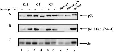
FIG. 7.
Effect of eIF4E induction on p70S6k phosphorylation. eIF4E-expressing cells were maintained in the presence or absence of tetracycline for 36 h (lanes 1 through 6); NIH 3T3 cells were grown in Dulbecco’s minimal essential medium containing 0.5% fetal bovine serum (FBS) for 16 h (lanes 7 to 9), after which cells were either mock treated (lane 7), incubated for 30 min in the presence of 20 ng/ml of rapamycin prior to stimulation with 10% FBS for 1 h (lane 8), or stimulated with 10% FBS alone (lane 9). Total cell extract (50 μg) was electrophoresed on two separate SDS–8% polyacrylamide gels and electroblotted onto a 0.45-μm-pore-size nitrocellulose membrane. One membrane was probed with a rabbit polyclonal anti-p70S6k antibody which recognizes p70S6k irrespective of its phosphorylation state (A), while the other was probed with a rabbit polyclonal anti-phosphopeptide antibody which is specific for phospho-Thr421 and phospho-Ser424 in p70S6k (B). (C) Total cell extract (20 μg) was immunoprecipitated with rabbit polyclonal anti-p70S6k antibody. The immunoprecipitate was assayed for p70S6k activity by using 40S ribosomal subunits as a substrate as described in Materials and Methods. The figure is a representative of three independent experiments.

