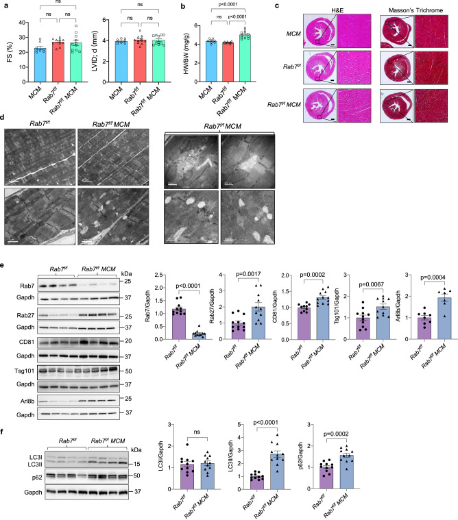
Fig. 6. Characterization of mice with cardiac specific deletion of Rab7.
a Echocardiographic analysis of ventricular function and structure at 28 days post-tamoxifen treatment. Percent (%) fractional shortening (FS) and left ventricular internal dimension in end diastole (LVID;d). MCM (n = 8 biologically independent animals), Rab7f/f (n = 11 biologically independent animals), Rab7f/f MCM (n = 11 biologically independent animals). b Heart weight to body weight ratio (HW/BW). MCM (n = 8), Rab7f/f (n = 11 biologically independent animals), Rab7f/f MCM (n = 11 biologically independent animals). c Hematoxylin and eosin (H&E) and Masson’s Trichrome staining of heart sections. Scale bar = 0.5 mm. d Ultrastructural analysis by transmission electron microscopy at D28 (representative of n = 2 biologically independent samples). e Representative Western blots and quantification of endosomal proteins (Rab7/Gapdh: Rab7f/f (n = 11 biologically independent animals) Rab7f/f MCM (n = 11 biologically independent animals); Rab27/Gapdh: Rab7f/f (n = 12 biologically independent animals), Rab7f/f MCM (n = 11 biologically independent animals); CD81/Gapdh: Rab7f/f (n = 10 biologically independent animals), Rab7f/f MCM (n = 12); Tsg101/Gapdh: Rab7f/f (n = 11 biologically independent animals), Rab7f/f MCM (n = 11 biologically independent animals); Arl8b/Gapdh: Rab7f/f (n = 8 biologically independent animals), Rab7f/f MCM (n = 7 biologically independent animals)). f Representative Western blots and quantification of autophagy proteins (n = 11 biologically independent animals). Data are mean ± SEM. ns = not significant. P values shown are by ANOVA with Tukey’s post-hoc testing (a) or two-sided Student’s t-test (e, f). Source data are provided as a Source Data file.

