Figure 5.
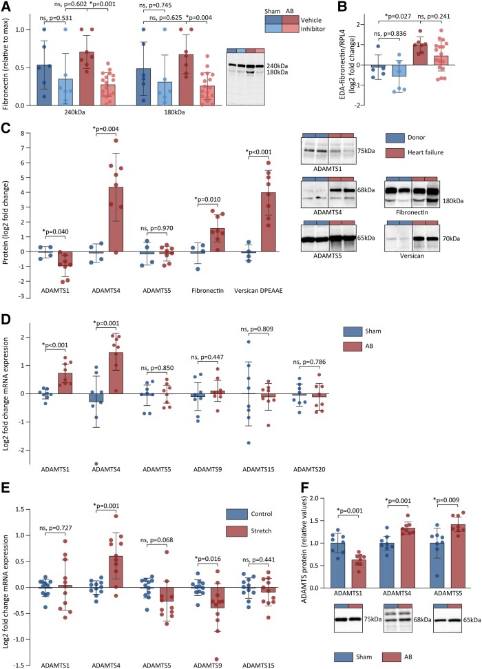
ADAMTS4 activity is increased in the myocardium of rats and patients with cardiac dysfunction. (A) The myocardial amount of full-length fibronectin (240 kDa) and its cleavage fragment (180 kDa) as determined by immunoblots in sham vehicle (n = 6), sham ADAMTS inhibitor (n = 6), AB vehicle (n = 7), and AB ADAMTS inhibitor (n = 17). Representative blots shown. (B) mRNA levels of EDA-fibronectin in myocardial samples from AB rats determined by RT–qPCR in sham vehicle (n = 5), sham ADAMTS inhibitor (n = 6), AB vehicle (n = 7), and AB ADAMTS inhibitor (n = 17). (C) The levels of active ADAMTS1, -4, and -5 proteins, fibronectin 180 kDa fragments, and versican DPEAAE fragments in myocardial samples from explanted human failing hearts (red) compared with healthy donor hearts (blue). Representative blots shown for ADAMTS4 (left), EDA-fibronectin (middle), and versican DPEAAE fragments (right). (D) mRNA levels of ADAMTS1, -4, -5, -9, -15, and -20 in myocardial samples from rats 6 weeks after AB (n = 8) or sham (n = 8). (E) Log2-transformed mRNA levels of ADAMTS1, -4, -5, -9, and -15 in adult human cardiac fibroblasts exposed to stretch (n = 11) compared to control conditions (n = 11). ADAMTS20 levels were not detectable. (F) The levels of ADAMTS1, -4, and -5 protein in myocardial lysates from rats 6 weeks after AB (n = 8) or sham (n = 8) relative to sham group. Bars represent mean ± 1 SD. Groups were compared by one-way ANOVA with planned comparisons followed by Bonferroni correction for the following comparisons: sham vehicle vs. sham ADAMTS inhibitor, sham vehicle vs. AB vehicle, and AB vehicle vs. AB ADAMTS inhibitor. Groups were compared by Student’s t-test for the following comparisons: sham vs. AB, control vs. stretched cells, and donor vs. heart failure patients. P < 0.05 were considered significant and marked with *. AB, aortic banding; EDA, extradomain A; ADAMTS4, a disintegrin and metalloprotease with thrombospondin motif; RT–qPCR, real-time quantitative polymerase chain reaction; DCM, dilated cardiomyopathy.

