
Fig. 2 |. Spatial localization and projection patterns of BFCNs.
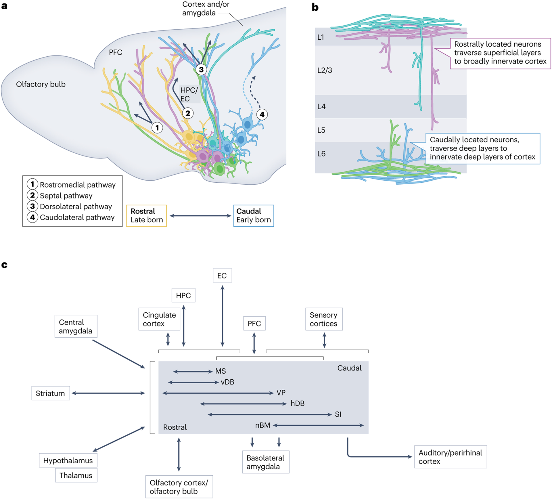
a, Overlapping pools of cholinergic neurons are located along the rostrocaudal extent of the basal forebrain in a manner that corresponds to their birthdate and site of origin162,216 (Fig. 1b,c). The axons of the basal forebrain cholinergic neurons (BFCNs) that make up these different rostrocaudal pools take four distinct projection paths to innervate their targets: the rostromedial pathway, the septal pathway, the dorsolateral pathway and the caudolateral pathway. In the schematic, arrows depict the relative trajectories of each of these fibre paths. Solid projections indicate fibres that traverse the sagittal plane, whereas those depicted with dashed lines are tracts that break from the sagittal plane and move laterally46,158. Developmentally diverse populations and their expected projection paths are shown in colours that correspond to their relative birth order (Fig. 1b,c). The major targets of each of the projections are noted. b, BFCNs innervate cortical layers distinctly on the basis of their rostrocaudal location in the basal forebrain46,158. Rostrally located, cortically projecting cholinergic neurons innervate both superficial and deeper layers of the cortex (purple and turquoise projection populations), whereas caudally located, cortically projecting cholinergic neurons primarily innervate deep layers of the cortex46,158 (blue and green). In both cases, fibre bundles traverse superficial or deep layers of the cortex as they find their targets46,158. c, An input and output wiring diagram for BFCN populations, derived from studies on the connectivity of cholinergic neurons34,64,144–176. The centre grey box organizes BFCNs beside horizontal arrows denoting their varying rostral (left) to caudal (right) extent. Regions shown to the left of the central BFCN box broadly innervate all BFCNs. Regions above and below the central BFCN box connect to specific BFCN population(s) denoted by the vertical arrows, highlighting approximate regional specificity in wiring. Brackets denote broad (rostrocaudal) overlap in connectivity across BFCN populations. The double-headed arrows denote reciprocal projections to a region, and the single-headed arrows denote unidirectional projections. EC, entorhinal cortex; hDB, horizontal subdivision of the diagonal band; HPC, hippocampus; L, layer; MS, medial septum; nBM, nucleus basalis of Meynert; PFC, prefrontal cortex, SI, substantia innominata; vDB, vertical diagonal band; VP, ventral pallidum.