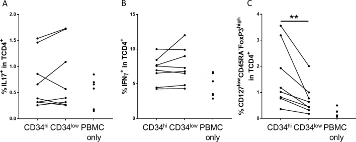
Figure 6.
CD4+ lymphocyte polarization, induced by CD34 sorted HUVECs. CD34high and CD34low HUVECs were seeded at a high confluence and after adhesion, they were co-cultured with PBMCs. Percentage of different CD4+ T lymphocytes, including Th17 (IL-17+ cells in CD4+T cells) (A), Th1 (IFNγ+ cells in CD4+T cells) (B), and Tregs (% CD127lowCD45RA−FoxP3high in CD4+T cells) (C) was evaluated at day 4 (C) and day 5 (A,B). PBMC alone (PBMC only) were used as a control. The results are shown as before and after representation and each dot correspond to an experiment (*p < 0.05, **p < 0.01: two-tailed Wilcoxon test).

