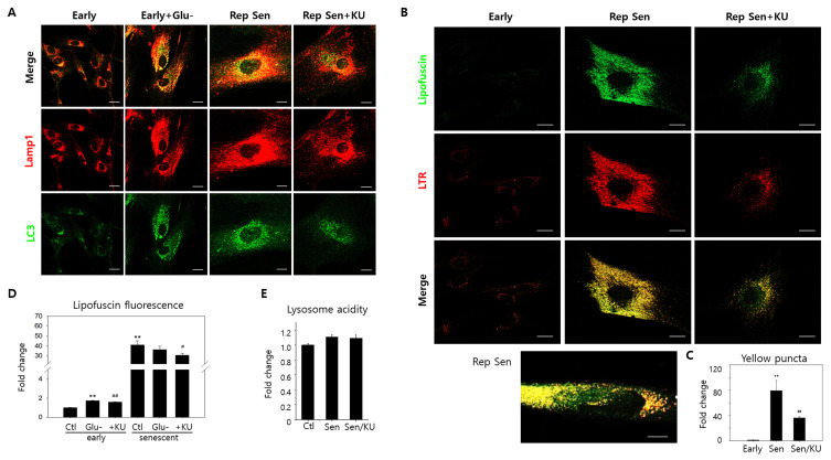
Fig. 2. Lipofuscin granule accumulation co-localized with autolysosomes in replicative senescence cells.
(A) Replicative senescence human fibroblasts (passage 31 or 32) (Rep Sen) were immune-stained with LC3 autophagosomes or Lamp1 lysosomes antibodies. Compared to early-passage cells (passage 20 or earlier) (Early), Lamp1 and LC3 positive puncta were far more abundant and co-localized (yellow puncta) with a pattern similar to glucose-starved cells (Early + Glu-). KU60019 treatment for three days lowered red and green puncta levels and co-localization. (B) As visualized through confocal microscopy, autofluorescent puncta levels substantially increased and were predominantly co-localized with lysosomes in senescent cells (Rep Sen). The bottom panel illustrates senescent cells presenting granular yellow puncta and cytosolic green fluorescence. KU60019 treatment attenuated lipofuscin granule and lysosome accumulation (Rep Sen + KU). LTR, LysoTrackRed. (C) Yellow puncta (over 0.5 μm2) were counted in ten sample cells visualized by confocal photographs, and normalized control cell values were plotted. (D) Early passage and replicative senescence cells were glucose-starved (Glu-) or treated with KU60019 (+KU). Flow cytometry examined 10,000 cells at 530 nm to determine cellular autofluorescence. Means normalized by the control were plotted (three biological repeats). (E) Early passage (Ctl) and senescent cells were either mock- (Sen) or KU60019 (Sen/KU)-treated. Lysosensor yellow/blue DND-160 dye stained 10,000 cells for lysosome acidity determination. ANOVA determined **P < 0.01, #P < 0.05, and ##P < 0.01 as significantly different. Scale bars = 20 μm.

