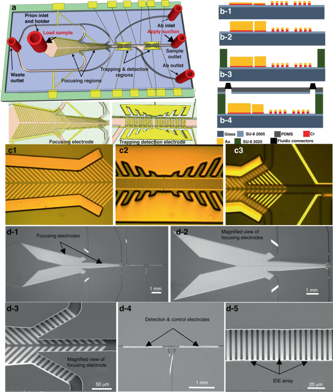
Fig. 1. Description of the Biosensor.
a 3-D view, b1–b4 sideview. c1–c3 Optical images of the biosensor after fabrication of the focusing electrode pair, the sensing and trapping electrodes array, and the SU8 microchannel. d1–d5 Scanning electron microscope (SEMs) micrographs of the fabricated biosensor. d-1 The two set focusing electrodes, detection electrode, and control electrode embedded in SU-8 microchannel, d-2 a magnified view of the two-set focusing electrode, d-3 magnified view of one focusing electrode, d-4 detection and control electrodes, d-5 a magnified view of the detection IDE array

