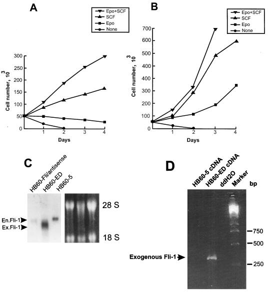
FIG. 3.
Effect of overexpression of Fli-1 on proliferation of HB60-5 cells by Epo. HB60-5 cells (5 × 10⁶) were transfected with either the sense or antisense CMV-Fli-1 expression vector (Fig. 8A), and the pools of transfected cells were incubated in the presence of growth factors as indicated. (A) Growth rate of the Fli-1 antisense-transfected HB60-5 cells. (B) Growth rate of the Epo-dependent HB60-ED cells, which are derived from the Fli-1 sense-transfected HB60-5 cells. (C) Analysis of expression of exogenous Fli-1 mRNA in transfected HB60-5 cells. mRNA extracted from HB60-5 cells, the pools of Fli-1 antisense-transfected HB60-5 cells, and HB60-ED cells were Northern blotted and hybridized with Fli-1 cDNA probe. The position of the exogenous Fli-1 (Ex.Fli-1) band, which is slightly smaller than endogenous Fli-1 (En.Fli-1) transcript, is shown by an arrowhead. The ethidium bromide-stained gel shows equal RNA loading. (D) Expression of the exogenous Fli-1 in HB60-ED cells was verified by PCR analysis using two primers corresponding to Fli-1 and transcription termination sequences from the pRc/CMV construct. The PCR products were separated on a 2% agarose gel and stained with ethidium bromide. The arrow shows the location of the 304-bp amplified fragment. A PCR using no cDNA(ddH2O) was used as a negative control.

