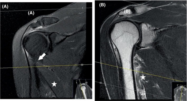
FIGURE 6.

Coronal proton density fatsat in a study (A) performed before the injury, showing a common placement of transverse slices in a regular shoulder MRI exam. The latissimus dorsi (star) and its proximal insertion (arrow) are not covered sufficiently by the axial slices (lines 1–22). Coronal T2 1 week post‐injury (B) was performed by an experienced radiographer who extended the imaging of axial slices to cover the injured area along the humerus.
