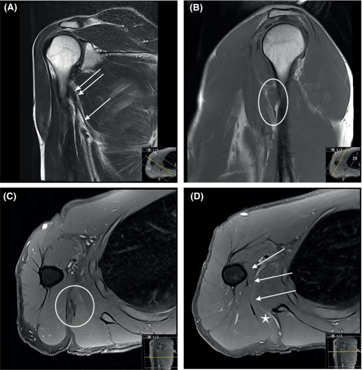
FIGURE 10.

Thirty‐eight weeks post‐injury. The hematoma and edema are completely absorbed. The muscle seems to have reattached to the humerus, and there is some residual scar tissue at the musculotendinous junction. (A) Coronal T2. The tendon can be seen as a slender structure all the way to its former insertion (arrows). (B) Sagittal T1. Part of the muscle is close to its humeral insertion indicated by the circle. Compare with previous Figure 7D. (C) Axial proton density (PD) fatsat. Scar tissue at the musculotendinous junction indicated by the circle. (D) Axial PD fatsat. The latissimus dorsi indicated by the star is reconnected to the humerus by reattachment of muscle tissue as indicated by the arrows.
