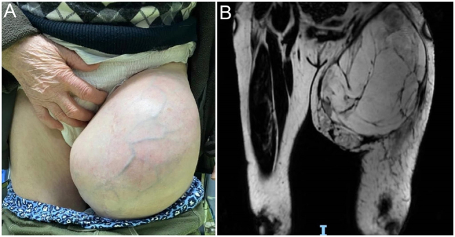
Figure 1.

A 77-year-old patient diagnosed approximately 2 years ago with liposarcoma is referred to our department with a rapidly increasing mass located at the root of the thigh (A). The MRI (coronal T1) revealed a tumor mass with a predominantly fatty structure, but inhomogeneous through gadolinophilic and cystic areas with contrast uptake at the level of walls and some septa, as well as left inguinal-femoral adenopathies (B).

 This work is licensed under a