Figure 2.
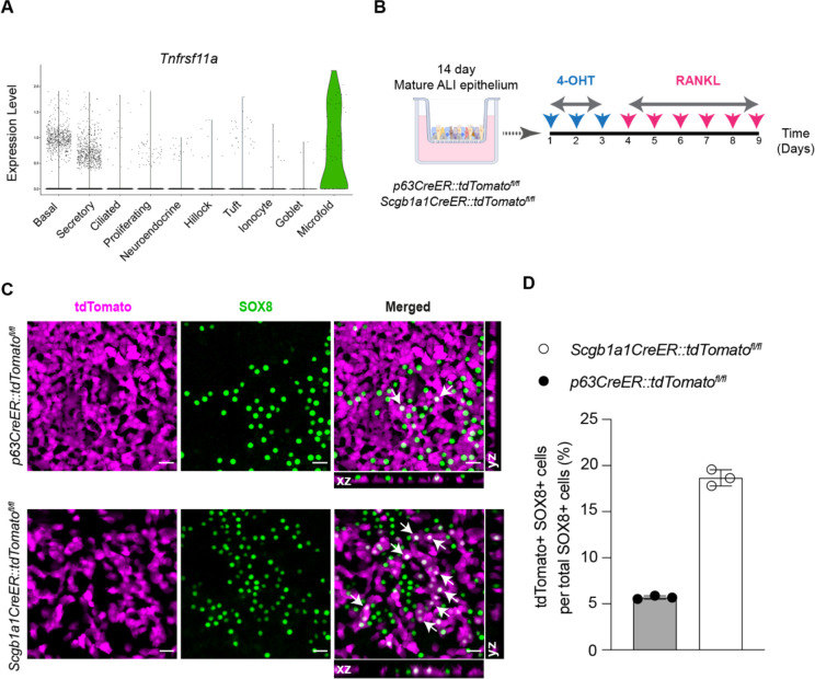
In vitro lineage tracing of airway M cells. (A) Violin plot of scRNA-seq profiles showing expression of Tnfrsf11a gene (RANK) in M cells as well as a small fraction of airway basal stem and secretory cells. (B) Schematic of in vitro lineage tracing of M cells in mouse tracheal ALI epithelial cultures derived from mice expressing cell-specific CreER drivers for basal stem (p63CreER) and secretory (Scgb1a1CreER) cells and the LSL-tdTomato reporter. 4-OHT stands for 4-hydroxytamoxifen. (C) Immunofluorescence images of p63CreER::tdTomatofl/fl and Scgb1a1CreER::tdTomatofl/fl lineage traced epithelial cells following RANKL-mediated M cell induction. Lineage positive M cells marked with arrows showing cytoplasmic tdTomato (magenta) and nuclear SOX8 (green) staining are seen in the merged panels and orthogonal sectional views. Scale bar, 20 μm. (D) Quantitative analysis of percent lineage positive M cells. Individual circles on the graph represent lineage positive cells in single RANKL-treated ALI epithelial culture wells. Data are presented as mean ± SD of triplicate ALI epithelial cultures.

