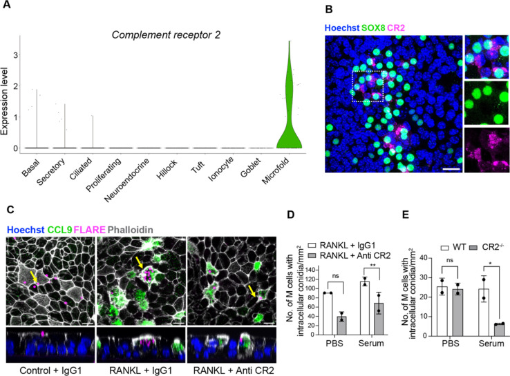
Figure 3.
Airway M cells express complement receptor 2 (CR2) and internalize Aspergillus fumigatus conidia in a CR2-dependent manner. (A) Violin plot showing specific expression of Cr2 in M cells based on scRNA-seq profiles. (B) Confocal micrograph of RANKL-induced mouse ALI epithelial cultures stained for M cell marker SOX8 (green) and CR2 (magenta) demonstrates CR2 expression in a subset of M cells. Boxed region is enlarged in insets on the right. Nuclei are stained with Hoechst dye (blue). Scale bar, 20 μm. (C) Confocal images showing FLARE conidia (magenta) adhering extracellularly to epithelial cells in control ALI (PBS) (left), internalization of multiple conidia inside CCL9 positive M cells (green) in RANKL-treated ALI (middle) and uptake of fewer conidia by M cells in RANKL-treated ALI pre-treated with Anti-CR2 antibody (Ab) (right). Epithelial cell membranes are marked by phalloidin (white). The panels below are orthogonal views (YZ) that include the cells marked by arrows in the respective panels above. Nuclei are stained with Hoechst dye (blue). Scale bar, 10 μm. (D) Quantification of endocytosed intra-M cell complement coated or uncoated FLARE conidia in RANKL-treated ALI cultures pre-treated with Control IgG1 or anti-CR2 antibodies. (E) Quantification of complement coated or uncoated FLARE conidia within M cells in RANKL-treated ALI cultures derived from wildtype (WT) or CR2−/− (CR2 knockout) basal cell lines. Individual circles on the graph represent intra-M cell conidia in single RANKL-treated ALI epithelial culture wells. Data are presented as mean ± SD of duplicate ALI cultures. ns = not significant, **P ≤ 0.01, and *P ≤ 0.05 by two-way ANOVA (Tukey’s multiple comparisons test).

