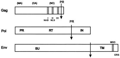
FIG. 3.
Details of the viral structural proteins. (Top) Gag protein. The locations of the three glycine-arginine-rich GR boxes (I, II, and III) are indicated by shaded boxes. The NAB and NLS are indicated below boxes I and II. The arrow indicates the cleavage site at which viral protease (PR) (encoded in Pol) cleaves about 3 kDa from the 78 kDa Gag precursor. The locations of the expected sites for the retroviral matrix (MA), capsid (CA), and nucleocapsid (NC) polypeptides are shown. These cleaved proteins, however, have not been found in mature, infectious virions. (Middle) Pol protein. The PR, RT, and integrase (IN) domains are shown. The PR cleavage site is indicated by the arrow labeled PR. (Bottom) Env protein. The surface (SU) and transmembrane (TM) domains are shown as well as the proteolytic cleavage site (arrow). MSD, membrane spanning domain in TM; ERS, endoplasmic reticulum sorting signal in the cytoplasmic tail.

