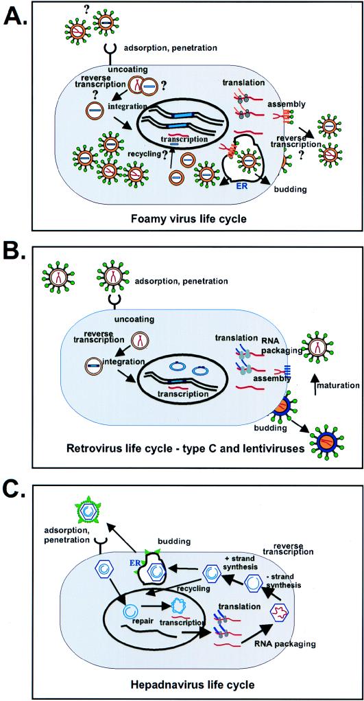
FIG. 4.
Replication pathway of FV compared to that of the conventional retroviruses and hepadnaviruses. (A) Proposed replication pathway of FVs. Viral RNA is indicated by red lines, and DNA is indicated by blue bars. Gold indicates the viral Gag protein, and green circles are the glycoproteins. Grey circles indicate polysomes. ER denotes endoplasmic reticulum. Several intracellular particles are shown, which represent the large numbers of intracellular particles in infected tissue culture cells which have been detected by both electron microscopy and viral assays. It is not known whether such particles contain RNA or DNA or both. The uncertain steps in the life cycle are indicated by question marks. (B) Retroviral life cycle. Colors used are the same as in panel A. Immature particles are shown with gold centers; after protease cleavage, the mature extracellular particles are shown with white central cores. Details of the replication pathway can be found in reference 16. (C) Hepadnavirus life cycle. Abbreviations are as in panels A and B. Viral core proteins are indicated by purple, RNA by red, DNA by blue, and S glycoproteins by green. Details of the replication pathway can be found in reference 25.

