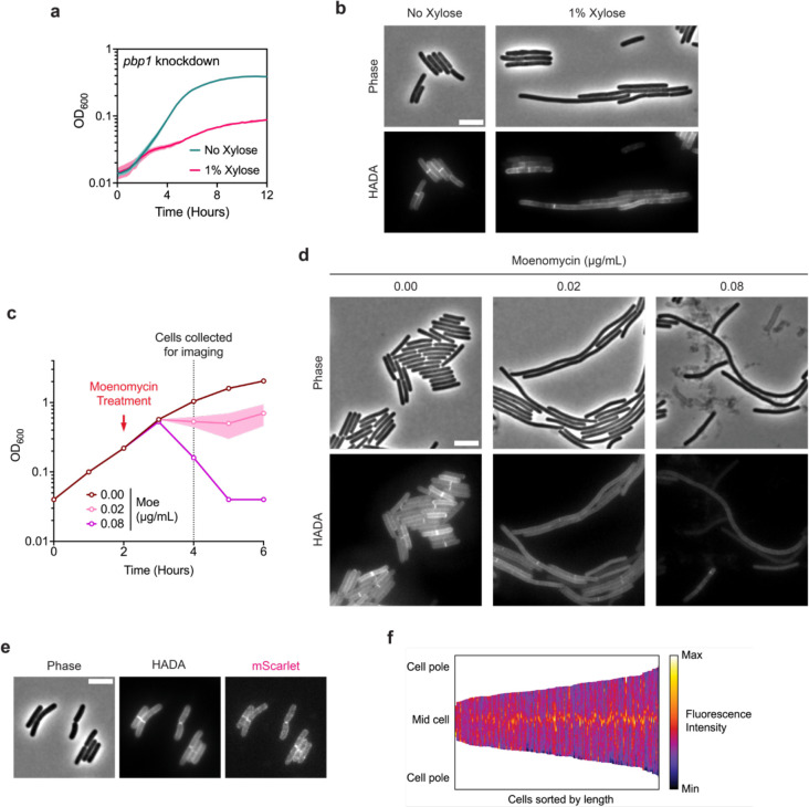
Fig. 5 |. The class A PBP, PBP1, is critical for cell division.
a, Growth profile of the CRISPRi pbp1 knockdown strain. Data from a single growth curve experiment; mean and standard deviation plotted from three biological replicates. b, Representative micrographs showing morphological and PG incorporation phenotypes of pbp1 knockdown cells. PG was labeled by incubation with HADA. Scale bar, 5 μm. Data representative of multiple experiments. c, Moenomycin treatment of WT cultures. Moenomycin was added after two hours of growth as indicated, and cells were collected and fixed for imaging 2 hours after treatment. Mean and range are plotted from two biological replicates. d, Representative micrographs showing morphological and PG incorporation phenotypes of moenomycin-treated WT cells. PG was labeled by incubation with HADA. Scale bar, 5 μm. e, Representative phase-contrast and fluorescence micrographs showing PG incorporation and subcellular localization of mScarlet-PBP1 in exponentially growing cells. PG was labeled by incubation with HADA, and mScarlet-pbp1 was expressed using the native pbp1 promoter from an ectopic locus. Scale bar, 5 μm. f, Demograph showing fluorescence intensity across multiple cells representing subcellular localization of PBP1 in exponentially growing cells. Cells are aligned at the mid-cell and sorted by length. Data are from 173 cells from one experiment and are representative of multiple experiments.

