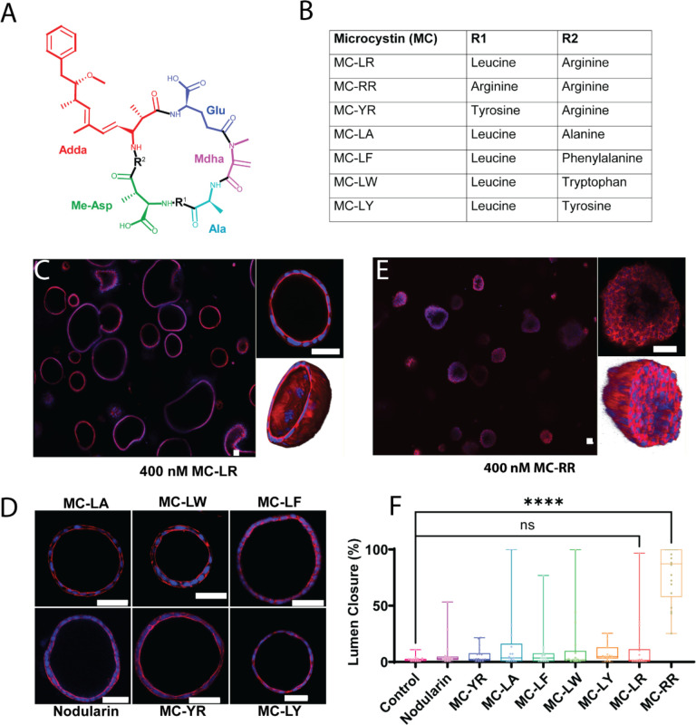
Figure 1: MC-RR treatment results in neonatal EHBD cholangiocyte spheroid damage.
A) General structure of microcystins showing cyclic heptapeptide backbone with two variable amino acid groups (marked as R1 and R2). Mdha: N-methyldehydroalanine, Adda: (all-S,all-E)-3-amino-9-methoxy-2,6,8-trimethyl-10-phenyldeca-4,6-dienoic acid, Me-Asp: D-erythro-β-methyl-isoaspartic acid, Ala: Alanine, Glu: Glutamic acid. B). Commercially available MCs tested, with identity of amino acids at R1 and R2 noted. C) Neonatal EHBD cholangiocyte spheroids forming one-cell thick hollow spheroids in presence of 400 nM MC-LR. The middle section of a representative spheroid is highlighted and the 3D rendering shows a typical hollow lumen. D) Neonatal EHBD cholangiocyte spheroids form one-cell thick hollow spheroids with filled lumens after treatment with 400 nM of various microcystins and nodularin for 24 h. E) Neonatal EHBD cholangiocyte spheroids forming multi-cell thick spheroids after treatment with 400 nM MC-RR for 24 h. The middle section of a representative spheroid is highlighted and the 3-D rendering shows the lumen filled with cells. F) Quantification of lumen closure in the presence of all the tested microcystins and nodularin. All images are representative of 3 independent experiments, with a minimum of 18 spheroids for each condition quantified for lumen closure. Control spheroids are vehicle treated. Red: actin, Blue: DAPI. All scale bars 50 μm.

