
Figure 4. Mode of m A binding by writer/sensor, eraser, and reader.
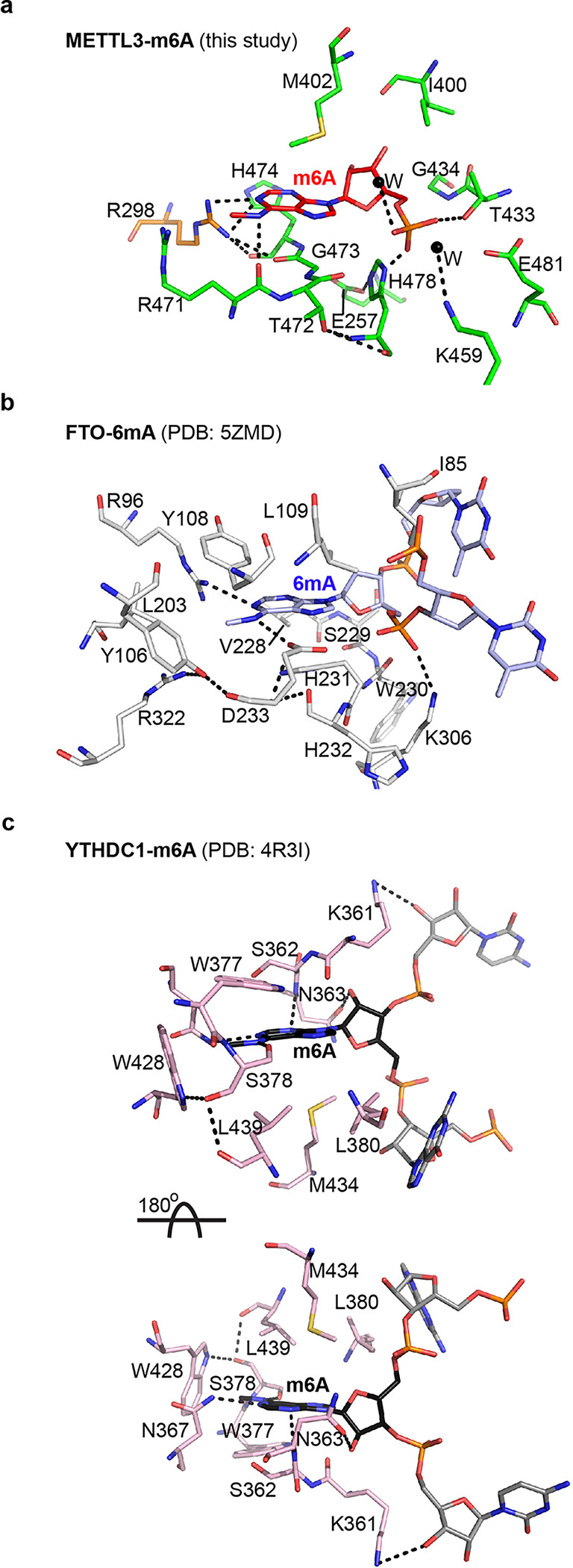
Interaction networks of m6A (red) binding to METTL3 (green), and METTL14 (a), 6mA (blue) binding to FTO (b), and m6A binding to YTH domain of YTHDC1 (c). The two nucleotides flanking the flipped methylated base in FTO and YTHDC1 are shown in light blue and grey, respectively. The hydrophobic stacking surface in YTHDC1 can only be aligned by rotating the molecule 180° around the x-axis, suggesting that reader proteins approach RNA from the opposite direction. The m6A pocket of METTL3-METTL14 harbors features that enable it to act as an atypical m6A sensor/reader during its switch from writer to reader. Dashed lines, h-bonds.