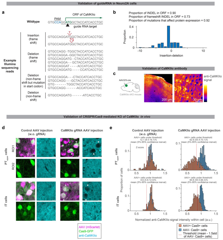
Extended Data Figure 3. Histological validation of CRISPR/Cas9 KO of CaMKIIα.
a. Sequencing of genomic DNA in Neuro2A cells transfected with guide RNA against CaMKIIα and Cas9 (Methods).
b. The proportion of amplicon with Indel mutation.
c. CaMKIIα immunohistochemistry of ALM in CaMKIIα conditional knock out (cKO) mouse injected with AAV-hsyn-cre. Loss of signal was observed around the injection site (ALM; the center of the image), validating the CaMKIIα antibody.
d. CaMKIIα immunohistochemistry of ALM with cell-type-specific CRISPR/Cas9 KO of CaMKIIα. Loss of CaMKIIα was observed only in cells expressing both Cas9 (green) and guide RNA (magenta, in right panels). Black areas smaller than PT/IT neurons are presumably glia, inhibitory neurons, or neuropile of manipulated cells.
e. Quantification of CaMKIIα immunostaining signal (Methods).

