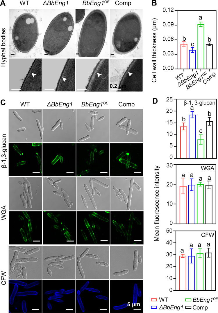
Fig 4. Cell wall structures of B. bassiana wild type and mutant strains.
A, B. TEM micrographs (scale: 0.2 μm) for cell walls of hyphal bodies and cell wall thickness (n = 30). C, D. Confocal microscopic images (scale: 5 μm) for cell wall β-1,3-glucan and chitin and measurements of their contents. β-1,3-glucan was labeled with monoclonal β-1,3-glucan specific antibody and goat anti-mouse IgG-FITC. Chitin was labeled with WGA or stained with CFW as detailed in the Methods section. Mean fluorescence intensities were quantified densitometrically using the ImageJ software. Error bars: SDs. Different letters indicate significant differences (P < 0.01 in LSD test).

